

Universitetsavisen
Nørregade 10
1165 København K
Tlf: 35 32 28 98 (mon-thurs)
E-mail: uni-avis@adm.ku.dk
—
Science
Medicine — Researchers have cured seven patients of a life-threatening skin disorder with a new method that finds defective proteins. Now they're looking at using the method on types of cancer.
The journal Nature Methods has named a new medical analysis method as the biggest scientific breakthrough of 2024.
But groundbreaking results achieved by the Mann Group, a research unit at the Faculty of Health and Medical Sciences at the University of Copenhagen (UCPH), may have been slightly overlooked.
It concerns a method that has led to a cure for patients suffering from a rare, but very unpleasant, skin disease Toxic Epidermal Necrolysis (TEN).
The disease causes the skin to form blisters and leads to the greyish peeling off of large areas of skin. It has a mortality rate of approximately 30 per cent. Many of the survivors also experience serious side effects like permanent deafness and blindness. Doctors had no medication against TEN until 2024.
But now the Mann Group has already cured seven patients of this rare skin disease using a completely new method. It has taken researchers a year to find a drug against the life-threatening disease.
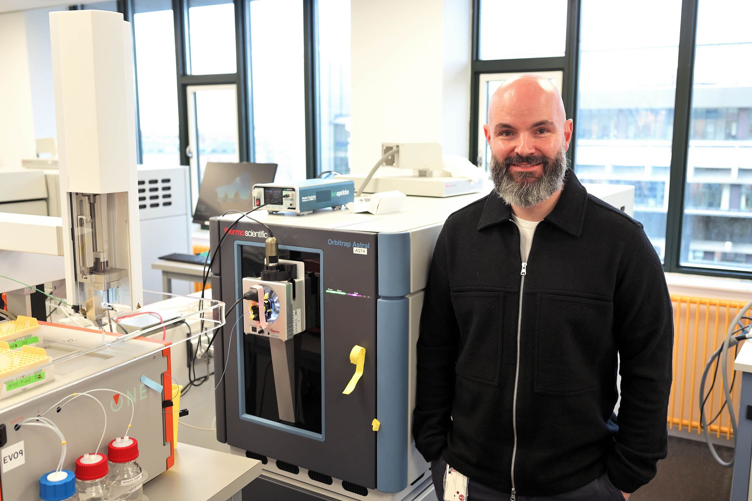
The Mann Group consists of 30 employees who work at the Novo Nordisk Foundation Center for Protein Research at the University of Copenhagen and at the Max Planck Institute of Biochemistry (MPIB) near Munich, Germany.
The idea for the new method came from Associate Professor Andreas Mund, who came to work in the University of Copenhagen’s health science Panum building complex for the first time in 2013 to work with microscopy on individual cells.
He joined the Mann Group in 2017, where he one day had an idea, he explains to the University Post when we visit him in his office. His idea depended on the ability to combine several different technologies:
It will change our understanding of tissue biology, human health and disease
Associate Professor Andreas Mund
»My manager, Matthias Mann, was open to my proposal from the very beginning, and the research group’s combined ability to collaborate and be innovative made it possible for me to turn my plan into action,« says Andreas Mund.
He is a graduate in biotechnology engineering and molecular biology from the University of Hamburg, but he considers himself first and foremost an engineer. He reckons that it is the engineering degree programme that gave him the opportunity to start the development of the new technology called DVP.
D is for deep learning, which includes the use of artificial intelligence; V is for visual, working with microscopy; and P is for proteomics, the science of the interactions, composition and structures of proteins and cells.
Proteomics is associated in DVP with the use of a mass spectrometer. It is the combination of proteomics and the mass spectrometer that lets you analyse different types of proteins in the cells in a tissue sample.
With DVP, the researchers in the Mann Group were suddenly able to see what differed, or what did not work, in the selected cells with proteins, in patients with TEN. This let them find a drug that corrects what the proteins in the cells do wrong — and which makes patients ill.
Back in 2017, Andreas Mund knew that his idea was on the edge of what was technically feasible. But as an engineer he had a good sense of how machines and technology are constantly evolving, and this gave him the courage to continue.
International collaboration
DVP technology has involved engineers, computer scientists, molecular biologists and pathologists from different countries.
The journal Nature Methods has named DVP as the research result of the year for 2024.
»It would not have been possible to get DVP to work just ten years ago. It wasn’t until 2018 that it became possible to build a mass spectrometer that could work with sufficient sensitivity. A mass spectrometer now has a sensitivity that corresponds to being able to register a fly settling on a jumbo jet,« says Andreas Mund.
On top of this is the use of artificial intelligence, which is also absolutely necessary in connection with DVP, but which has only been developed in recent years.
Today, almost half of the Mann Group staff work on developing software programmes and AI that can process the large amounts of data obtained from analysing tissue samples with hundreds of thousands of cells, and even more proteins.
But when Andreas Mund joined the Mann Group in 2017, he could see that it would be possible to combine what he calls the best parts of image analysis with the best parts of proteomics.
There is one more technology that has required a lot of development in order to be applicable in DVP.
Since 2018, Mann Group has been working with Leica, one of the world’s leading manufacturers of lenses and microscopes, to develop a technology that can cut out a cell out with a laser beam, according to Andreas Mund.
To be able to assess whether the proteins in a cell are behaving right, the researchers place a tissue sample in formalin and then in paraffin, so that it is possible to cut it into slices with a thickness of 3-5 micrometres (thousandths of a millimetre), which can then be examined under a microscope.
We are now working on studying different types of cancer, infections caused by viruses, as well as arteriosclerosis and liver diseases
Associate Professor Andreas Mund
What’s new is that in the Mann Group they can now set up a digital map in an extremely high resolution for the entire sample using AI, so they can identify each of the hundreds of thousands of cells.
»We give each cell an x-y coordinate, which we send to another microscope that, using a laser beam, can extract the cells one by one if we suspect that they contain disease and important information about a cell’s proteins. The Al technology has been developed here on the sixth floor of the Panum complex, together with the Mann Group at MPIP in Germany and in collaboration with a Hungarian expert in image analysis with AI,« says Andreas Mund.
Many parties are involved now, but the idea of DVP originated with Andreas Mund, who says that he had learned to study one cell at a time:
»One day in 2017, I saw a picture of several cells and their phenotypes. I stood there looking at it and thought, what do you do now? I had learned about proteomics in the Mann Group, and it suddenly struck me that it seemed obvious to combine high-resolution microscopy with proteomics,« he says and adds:
»It will change our understanding of tissue biology, human health, and disease.«
According to Andreas Mund, other research groups around the world are starting to use the DVP technology, but things take time.
»It is still difficult to work with DVP and get it to work in practice. Many research groups are interested though, and more laboratories have started to join the effort. In addition, we at the Mann Group are working to spread the technology out to as many as possible,« he says.
Mann Group also focuses on several other diseases in which proteins play an important role, according to Andreas Mund:
»We are now working on studying different types of cancer, infections from viruses, as well as arteriosclerosis and liver diseases, and our hope is that we will understand these diseases so well that we can find a cure for them«.